Contacto
Barranca del Muerto 520, Los Alpes, Álvaro Obregón, C.P.01010, Ciudad de México, MéxicoTeléfono
(+52) 55 9171 9570
Irène Joliot-Curie (1897–1956), hija de Pierre y Marie Curie, fue la segunda mujer en su familia en recibir el Nobel. Junto a su esposo Frédéric descubrió la radiactividad inducida en 1935. Durante la I Guerra Mundial trabajó como enfermera radiológica en el frente. Doctora en física, investigadora del Instituto del Radium y feminista activa, fue también una de las primeras mujeres en el gobierno francés.
Irène Joliot-Curie (1897–1956), daughter of Pierre and Marie Curie, became the second woman in her family to win a Nobel Prize. With her husband Frédéric, she discovered induced radioactivity in 1935. During WWI she served as a radiology nurse at the front. A physics PhD, researcher at the Radium Institute, and committed feminist, she was also among the first women to serve in the French government.
Irène Joliot-Curie (1897–1956), filha de Pierre e Marie Curie, foi a segunda mulher de sua família a receber o Nobel. Com o marido Frédéric, descobriu a radioatividade induzida em 1935. Durante a Primeira Guerra Mundial atuou como enfermeira radiológica na linha de frente. Doutora em física, pesquisadora do Instituto do Rádio e feminista engajada, foi também uma das primeiras mulheres a integrar o governo francês.
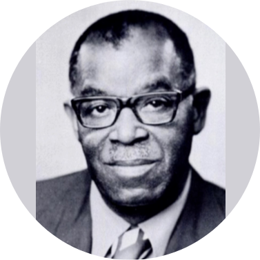
William J. Allen (1903–1981), primer radiólogo afroamericano certificado por el American Board of Radiology. Graduado en Howard University, combinó medicina y música. En el Homer G. Phillips Hospital creó un programa de residencia que formó a más de 200 técnicos de color. Teniente coronel en la II Guerra Mundial, lideró radiología en Fort Huachuca y fundó la sección de radiología de la NMA. Su legado abrió caminos y oportunidades para su comunidad.
William J. Allen (1903–1981), first African American radiologist certified by the American Board of Radiology. Graduate of Howard University, he combined medicine with his love for music. At Homer G. Phillips Hospital, he created a residency program that trained over 200 Black technicians. A WWII lieutenant colonel, he led radiology at Fort Huachuca and founded the NMA radiology section. His legacy broke barriers, expanded opportunities, and inspired generations.
William J. Allen (1903–1981), primeiro radiologista afro-americano certificado pelo American Board of Radiology. Formado pela Howard University, uniu medicina e música. No Hospital Homer G. Phillips, criou um programa de residência que formou mais de 200 técnicos negros. Tenente-coronel na Segunda Guerra Mundial, liderou radiologia em Fort Huachuca e fundou a seção de radiologia da NMA. Seu legado rompeu barreiras, ampliou oportunidades e inspirou gerações.
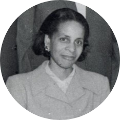
Ivy, pionera afroamericana en radiología, nació en Brooklyn en 1916. Tras iniciar su carrera en nutrición y servir como teniente en la II Guerra Mundial, estudió medicina en Meharry y se especializó en radiología en Tuskegee, donde fue directora hasta 1986. Reconocida por su liderazgo, obtuvo premios nacionales y dejó un legado de superación frente al racismo y sexismo, inspirando a futuras generaciones médicas.
Ivy, African American pioneer in radiology, born in Brooklyn in 1916. After a career in nutrition and service as a lieutenant during WWII, she studied medicine at Meharry and specialized in radiology at Tuskegee, where she became director until 1986. Honored nationally, she overcame racism and sexism, leaving a legacy of resilience and inspiration for future generations of women in medicine.
Ivy, pioneira afro-americana em radiologia, nascida no Brooklyn em 1916. Após carreira em nutrição e serviço como tenente na Segunda Guerra Mundial, estudou medicina em Meharry e se especializou em radiologia em Tuskegee, onde foi diretora até 1986. Reconhecida nacionalmente, superou racismo e sexismo, deixando um legado de perseverança e inspiração para futuras gerações de mulheres na medicina.
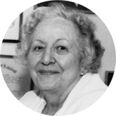
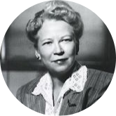
Lucy Squire fue una radióloga americana (1915-1996) y primera mujer residente en radiología del Massachusetts General Hospital de Boston. Nacida en Washington de padres canadiense y alemán-americano, estudió medicina en la George Washington School of Medicine y Woman's Medical College, graduándose en 1940. Pionera en educación radiológica, escribió "Fundamentals in Radiology" en 1964, que se convirtió en referencia mundial. En 1987 recibió el primer Marie Curie Award de la American Association of Women Radiologists.
Lucy Squire was an American radiologist (1915-1996) and the first female resident in radiology at Massachusetts General Hospital in Boston. Born in Washington to Canadian and German-American parents, she studied medicine at George Washington School of Medicine and Woman's Medical College, graduating in 1940. A pioneer in radiological education, she wrote "Fundamentals in Radiology" in 1964, which became a worldwide reference. In 1987 she received the first Marie Curie Award from the American Association of Women Radiologists.
Lucy Squire foi uma radiologista americana (1915-1996) e a primeira mulher residente em radiologia do Massachusetts General Hospital em Boston. Nascida em Washington de pais canadense e germano-americano, estudou medicina na George Washington School of Medicine e Woman's Medical College, formando-se em 1940. Pioneira em educação radiológica, escreveu "Fundamentals in Radiology" em 1964, que se tornou uma referência mundial. Em 1987 recebeu o primeiro Marie Curie Award da American Association of Women Radiologists.
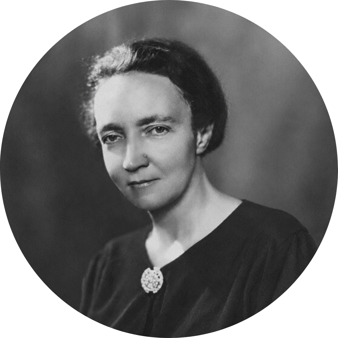
Edith H. Quimby (1891-1982) fue una pionera en medicina nuclear y física, destacándose en la protección contra radiaciones ionizantes. Nacida en Rockford, Illinois, fue la primera mujer en recibir la Medalla Janeway y desempeñó roles clave en diversas instituciones, incluyendo Cornell y Columbia. Su investigación definió límites seguros de radiación para tratamientos médicos, influyendo en la dosificación para cáncer. Reconocida por su contribución al campo, su legado perdura con el Edith H. Quimby Lifetime Achievement Award.
Edith H. Quimby (1891-1982) was a pioneer in nuclear medicine and physics, excelling in protection against ionizing radiation. Born in Rockford, Illinois, she was the first woman to receive the Janeway Medal and held key roles at various institutions, including Cornell and Columbia. Her research defined safe radiation limits for medical treatments, influencing dosages for cancer. Recognized for her contributions to the field, her legacy endures with the Edith H. Quimby Lifetime Achievement Award.
Edith H. Quimby (1891-1982) foi uma pioneira em medicina nuclear e física, destacando-se na proteção contra radiações ionizantes. Nascida em Rockford, Illinois, foi a primeira mulher a receber a Medalha Janeway e ocupou papéis importantes em várias instituições, incluindo Cornell e Columbia. Sua pesquisa definiu limites seguros de radiação para tratamentos médicos, influenciando a dosagem para câncer. Reconhecida por suas contribuições ao campo, seu legado perdura com o Prêmio Edith H. Quimby de Realização Vitalícia.